Essential Eye Exam Equipment
Modern-day eye care is filled with exciting, cutting-edge equipment for the treatment and diagnosis of various eye conditions. From ultra-widefield Optos cameras that can capture extensive views of the peripheral retina, to advancements in Optical Coherence Tomography that can image, non-invasively, high-definition, real-time, cross-sections of ocular tissue, to laser-assisted cataract surgeries, modern eye care touts earlier disease detection and improved treatment outcomes. However, these tools are often expensive, require a consistent electrical supply, and, more often than not, are designed for patients without any disability. In parts of the world where cutting-edge eye clinics are not feasible and for clinics serving a large special-needs population, let's go back to the basics, hone our clinical skills, and maximize our resources to deliver quality and versatile eye care worldwide. Below are my recommendations for outfitting a highly accessible eye clinic:
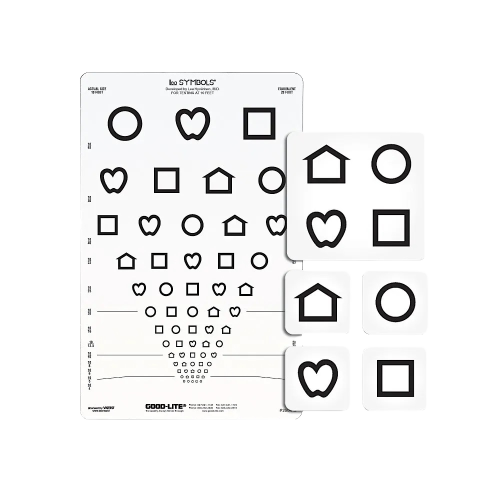
LEA Eye Chart
an optotype developed by Finnish ophthalmologist, Lea Hyvärinen enables visual acuity testing for patients with various communication needs: + simple point-and-match instructions + literacy not required + works well in all clinic settings
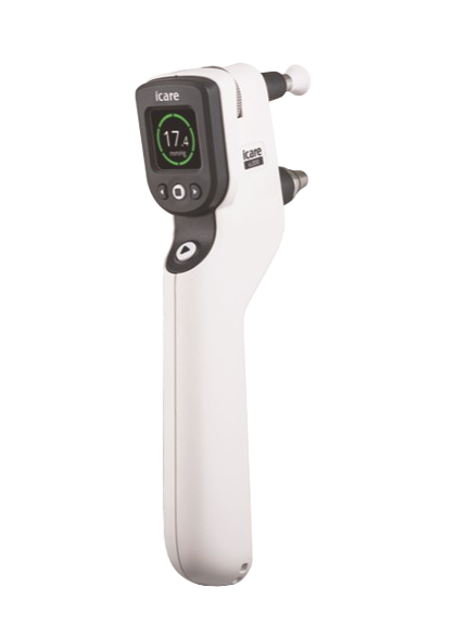
Rebound Tonometer
This is the one area where I recommend adopting the latest cutting-edge technology. The iCare IC 200 rebound tonometer, developed by a Finnish ophthalmic company, Revenio Group, single-handedly addressed all the pain points of previous IOP measurement tools. + battery operated + ultraportable + disposable probes + no anesthetic drop or fluoresceine dye required + easy to operate/train a technician + accurate IOP measurements + extensive flexibility in patient positioning (works in 200° measurement angles) accommodates bed-bound patients in supine positions
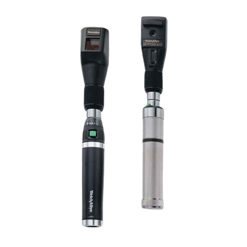
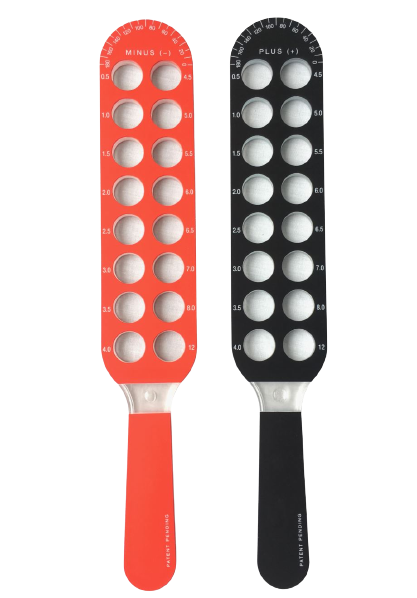
Traditional Retinoscope and Ret Racks a.k.a. Skiascopy Bars
provides an ultraportable, objective diagnosis of refractive errors + compact, easy to transport + quick and effective + retinoscope can be rechargeable or battery operated depending on electrical needs - requires significant training to perfect the techniques
Click Here for a Helpful Retinoscopy Simulator Helpful Videos on Retinoscopy:
Refraction Tools
Refining refractions can be streamlined using:
1. Traditional Phoropters mounted on a tripod or stand: + quick & efficient - bulky to transport
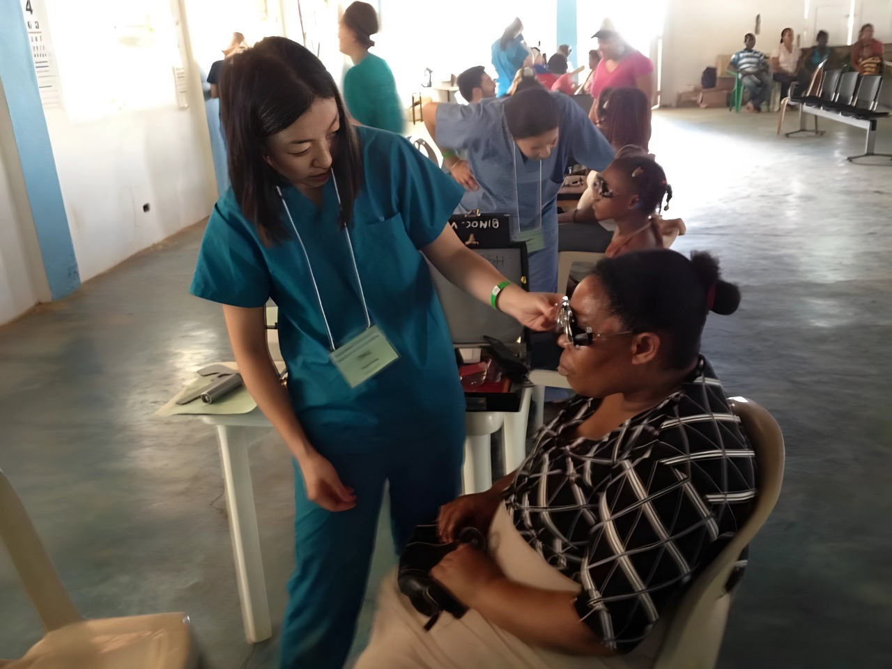
2. Trial Frame and Loose Lenses: + preview of glasses prescription + easy to transport - refractions may take a little longer
Direct Ophthalmoscope and 20D Condensing Lens
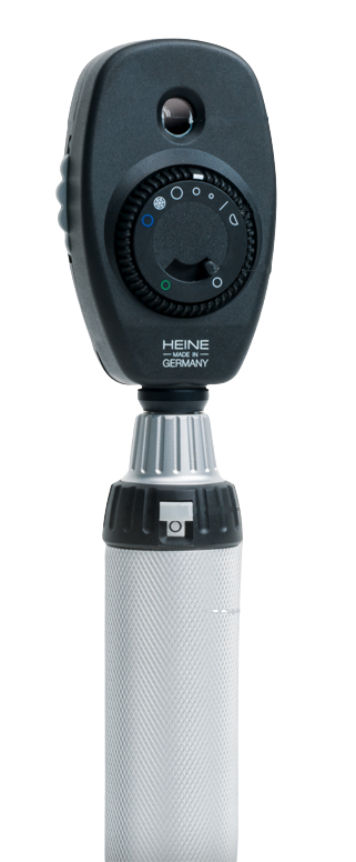
1. Using an ophthalmoscope on the slit beam setting and adjusting the working distance of a 20D lens produces quality views of the anterior segment structures The slit aperture is available on most ophthalmoscope models (Heine, Welch Allyn, and most Keeler models except for the Standard Keeler O-scope model)
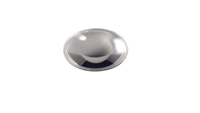
2. Holding the O-scope between the examiner’s eyes and the 20D lens at the appropriate working distance from the patient’s pupil is an adequate replacement for the traditional binocular indirect ophthalmoscope (BIO) headset for fundus examination
